Innovations in MPI
Innovations in MPI
Technological Advances That May Benefit Your Lab and Patients
Nuclear cardiology has rapidly evolved over the years. Technological innovations in hardware and software have helped improve imaging quality and diagnostic accuracy, and reduce radiotracer exposure.1
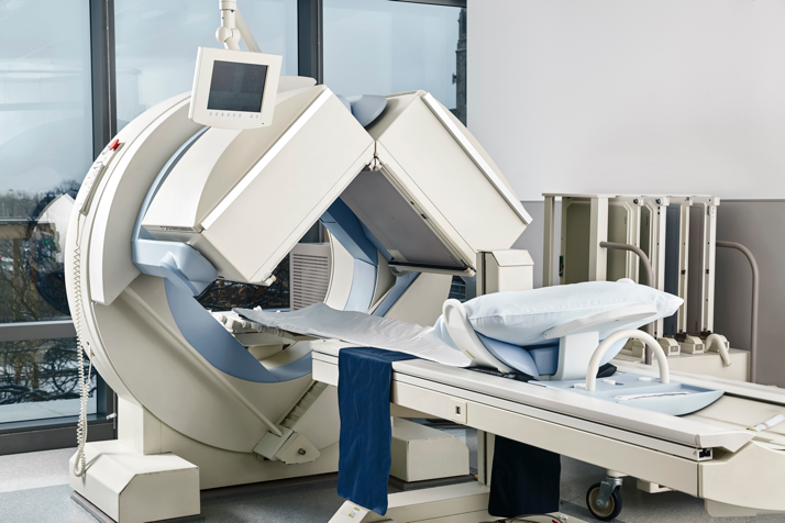
SPECT remains the most commonly used modality for the evaluation and risk stratification of patients with known or suspected CAD, partly because it is widely available, relatively less expensive than other modalities, and is compatible with a wider variety of radiopharmaceuticals when compared to PET.1,2
- Increased photon sensitivity through the addition of high-sensitivity collimation and multiple detectors1
- Improved energy resolution with today’s solid-state detectors made of CZT, which has up to 10 times higher sensitivity than conventional gamma cameras3
- Allowed for reduced radiotracer dose and radiation exposure, and decreased acquisition time1

- Improved spatial resolution
- Reduced scan time by half to a quarter
- Improved image quality
Advances in stress imaging protocols, camera technology, and processing software can enhance diagnostic accuracy and reduce radiation exposure.5 More advanced techniques help reduce motion artifacts and potentially improve the patient experience due to reduced imaging times.5 In addition, because contemporary SPECT cameras have made the acquisition of dynamic SPECT easier, quantification of myocardial blood and flow reserve is now possible to detect multivessel CAD.6
There have also been several advances in PET instrumentation that have improved various aspects of imaging. PET sensitivity has been improved with a larger axial field of view and 3D imaging. Advances have also been made to reconstruction software, which integrates resolution recovery with advanced iterative techniques.7
The cardiac PET radiopharmaceuticals have a higher extraction fraction compared to SPECT, which allows for better tracking of blood flow at higher rates produced by pharmacologic stress. This leads to an increase in the detection of moderate-severity CAD.8

Hybrid imaging systems that combine nuclear imaging with computed tomography (CT) allow for combined anatomic and functional testing.9 The combination also allows physiological assessment of flow and the anatomic extent and severity of CAD, while providing information about plaque composition and arterial remodeling.9 In addition, CT offers the ability for coronary calcium assessment, which adds prognostic value.1
MPI studies combined with other noninvasive measures such as myocardial blood flow (MBF) and coronary artery calcium (CAC) score have gained attention for potentially providing incremental diagnostic and prognostic value and impacting clinical decision-making.5,10
While the diagnosis of CAD has traditionally focused on obstructive atherosclerosis of the arteries, focus has turned to functional and structural abnormalities of the microvasculature.11 MBF and myocardial flow reserve (MFR) are measurements of the functional status of the coronary microvasculature that can10:
- Detect microvascular dysfunction
- Facilitate the diagnosis of multivessel CAD
- Describe changes between 2 studies in the same patient
MBF is the absolute measure of blood flow in units of mL/min/g.6 MFR is the ratio of MBF at stress to MBF at rest. According to the ASNC guidelines, MBF/MFR is most useful for8:
- Patients with no prior history of cardiac disease who present with symptoms of myocardial ischemia
- Patients with known CAD who may need more specific physiological assessment
- Identifying suspected multivessel CAD
- Assessing microvascular dysfunction when there is a disparity between visual perfusion abnormalities and normal coronary angiography
- Heart transplant patients with potential vascular abnormalities
Although somewhat empirical, ASNC Imaging Guidelines suggest that MFR values >2.3 indicate a favorable prognosis, and values <1.5 suggest significantly diminished flow reserve and elevated cardiac risk.10
PET MBF/MFR is an established acquisition approach that has been validated in clinical studies. Newer nonrotating SPECT systems have made absolute SPECT MBF/MFR quantification possible; this is being investigated to assess its incremental value in the diagnosis and risk stratification of patients with known or suspected CAD.6
While nuclear MPI provides a functional assessment of the extent, severity, and location of perfusion defects, anatomical assessment from CAC scoring can help identify patients at risk for CAD and help predict future coronary events. Used in combination with MPI, CAC scoring can add incremental prognostic value and help accurately identify patients at risk of CAD.5,12
The combination of stress MPI and CAC scoring allows the assessment of nonobstructive as well as obstructive CAD. Results from both anatomical and functional assessments may improve diagnostic accuracy and better define patient risk.12
FFR derived from computed tomography angiography (CTA) (FFR-CT) allows noninvasive assessment of hemodynamically significant lesions in addition to the anatomical assessment of coronary stenosis.13
A 2020 study suggests a relationship between FFR-CT results and risk of all-cause death or myocardial infarction (MI). The study included 4737 patients from the ADVANCE (Assessing Diagnostic Value of Non-invasive FFR-CT in Coronary Care) Registry to evaluate the relationship between FFR-CT and clinical outcomes at 1 year. The patients had all been evaluated for suspected CAD and had atherosclerosis identified by CTA and coronary CTA submitted for FFR-CT analysis. Data trends were as follows.13
- All-cause death or MI occurred in 1.2% of patients with an FFR-CT ≤0.80 compared with 0.6% of patients with FFR-CT >0.80 (P=0.06)
- Patients who experienced a major adverse cardiac event (MACE) had lower mean FFR-CT values compared with those who did not have a MACE (0.70 ± 0.13 vs 0.74 ± 0.12; P=0.02)
- Time to first cardiovascular death or MI was significantly more common in patients with FFR-CT ≤0.80 than those with FFR-CT >0.80 (0.80% vs 0.20%; P=0.01)
- Abnormal FFR-CT values (≤0.80) were associated with more revascularization compared with normal FFR-CT values (>0.80) (38.4% vs 5.6%, P<0.001)
Diagnostic Value of Noninvasive FFR-CT13


References+
1. Abbott BG, Case JA, Dorbala S, et al. Contemporary cardiac SPECT imaging—innovations and best practices: an information statement from the American Society of Nuclear Cardiology. J Nucl Cardiol 2018;25(5):1847-60. 2. Li Z, Gupte AA, Zhang A, Hamilton DJ. PET imaging and its application in cardiovascular diseases. Methodist Debakey Cardiovasc J 2017;13(1):29-33. 3. Lee WW. Recent advances in nuclear cardiology. Nucl Med Mol Imaging 2016;50(3):196-206. 4. DePuey EG, Bommireddipalli S, Clark J, Thompson L, Srour Y. Wide beam reconstruction "quarter-time" gated myocardial perfusion SPECT functional imaging: a comparison to "full-time" ordered subset expectation maximum. J Nucl Cardiol 2009;16(5):736-52. 5. Dorbala S, Di Carli MF. Nuclear cardiology. In: Libby P, Bonow RO, Mann DL, Tomaselli GF, Bhatt DL, Solomon SD, eds. Braunwald’s Heart Disease: A Textbook of Cardiovascular Medicine. 12th ed. Philadelphia, PA: Elsevier Inc, 2022. 6. Dorbala S, Ananthasubramaniam K, Armstrong IS, et al. Single photon emission computed tomography (SPECT) myocardial perfusion imaging guidelines: instrumentation, acquisition, processing, and interpretation. J Nucl Cardiol 2018;25(5):1784-846. 7. Slomka PJ, Pan T, Germano G. Recent advances and future progress in PET instrumentation. Semin Nucl Med 2016;46(1):5-19. 8. Bateman TM, Heller GV, McGhie AI, et al. Diagnostic accuracy of rest/stress ECG-gated Rb-82 myocardial perfusion PET: Comparison with ECG-gated Tc-99m sestamibi SPECT. J Nucl Cardiol 2006;13(1):24-33. 9. Fihn SD, Gardin JM, Abrams J, et al. 2012 ACCF/AHA/ACP/AATS/PCNA/SCAI/STS guideline for the diagnosis and management of patients with stable ischemic heart disease. J Am Coll Cardiol 2012;60(24):e44-164. Erratum in: J Am Coll Cardiol 2014;63(15):1588-90. 10. Dilsizian V, Bacharach SL, Beanlands RS, et al. ASNC imaging guidelines/SNMMI procedure standard for positron emission tomography (PET) nuclear cardiology procedures. J Nucl Cardiol 2016;23(5):1187-226. 11. Ziadi MC. Myocardial flow reserve (MFR) with positron emission tomography (PET)/computed tomography (CT): clinical impact in diagnosis and prognosis. Cardiovasc Diagn Ther 2017;7(2):206-18. 12. Engbers EM, Timmer JR, Ottervanger JP, Mouden M, Knollema S, Jager PL. Prognostic value of coronary artery calcium scoring in addition to single-photon emission computed tomographic myocardial perfusion imaging in symptomatic patients. Circ Cardiovasc Imaging 2016;9(5):e003966. 13. Patel MR, Nørgaard BL, Fairbairn TA, et al. 1-year impact on medical practice and clinical outcomes of FFRCT. JACC Cardiovasc Imaging 2020;13(1):97-105.